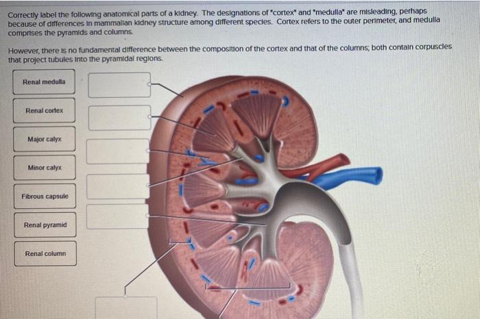
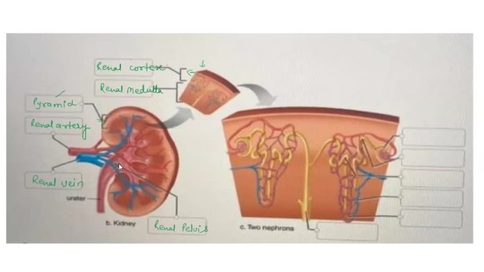
Correctly label the following anatomical parts of a kidney – Correctly labeling the anatomical parts of a kidney is crucial for accurate medical communication, enhanced understanding of kidney anatomy, and reduced risk of medical errors. This guide provides a comprehensive overview of the methods, procedures, and benefits of labeling kidney anatomy.
Understanding the precise location and function of each anatomical part is essential for accurate diagnosis and treatment of kidney-related conditions.
Introduction

The kidney is a vital organ responsible for filtering waste products from the blood and producing urine. Accurately labeling its anatomical parts is essential for effective communication among healthcare professionals and enhanced understanding of kidney function.
Methods for Labeling Anatomical Parts of the Kidney

Using Anatomical Diagrams
Anatomical diagrams provide visual representations of the kidney’s structures, allowing for easy identification and labeling.
Using Interactive 3D Models, Correctly label the following anatomical parts of a kidney
Interactive 3D models offer a dynamic way to explore the kidney’s anatomy, enabling users to rotate and zoom in for detailed labeling.
Using Textbooks or Online Resources
Textbooks and online resources provide detailed descriptions and images of the kidney’s anatomy, facilitating accurate labeling.
Examples of Anatomical Parts of the Kidney: Correctly Label The Following Anatomical Parts Of A Kidney

Renal Cortex
The renal cortex is the outermost layer of the kidney, responsible for filtering blood and producing urine.
Renal Medulla
The renal medulla is the inner layer of the kidney, involved in the concentration and modification of urine.
Renal Pelvis
The renal pelvis is the funnel-shaped structure that collects urine from the kidney’s calyces.
Ureter
The ureter is the tube that carries urine from the kidney to the bladder.
Procedures for Labeling Anatomical Parts of the Kidney
Identifying the Different Anatomical Parts
Carefully examine the kidney’s structure to identify its different anatomical parts.
Using the Correct Terminology
Use the standard anatomical terminology to accurately label the kidney’s parts.
Placing Labels in the Correct Location
Ensure that labels are placed in the correct anatomical location to avoid confusion.
Benefits of Correctly Labeling Anatomical Parts of the Kidney
Improved Communication among Healthcare Professionals
Accurate labeling enables clear and effective communication about kidney anatomy and pathology.
Enhanced Understanding of Kidney Anatomy
Correct labeling aids in understanding the structure and function of the kidney.
Reduced Risk of Medical Errors
Proper labeling minimizes the risk of misidentification and subsequent medical errors.
Quick FAQs
Why is it important to correctly label anatomical parts of the kidney?
Correct labeling ensures accurate communication among healthcare professionals, enhances understanding of kidney anatomy, and reduces the risk of medical errors.
What are the different methods used to label anatomical parts of the kidney?
Anatomical diagrams, interactive 3D models, textbooks, and online resources can be used for labeling kidney anatomy.
What are some examples of anatomical parts of the kidney?
Renal cortex, renal medulla, renal pelvis, and ureter are examples of anatomical parts of the kidney.
What are the benefits of correctly labeling anatomical parts of the kidney?
Correct labeling improves communication, enhances understanding of kidney anatomy, and reduces the risk of medical errors.